Monkey see Monkey do

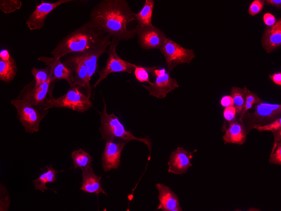
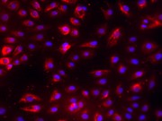
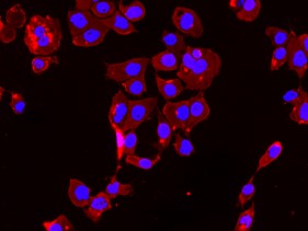
Last year, Zhen Liu et al., from the Institute of Neuroscience of the Chinese Academy of Sciences cloned Macaque monkeys (Macaca fascicularis), by somatic cell nuclear transfer (SCNT), the same cloning technique used to produce Dolly the sheep.


Cloning of animals by SCNT has been achieved in 23 mammalian species, the first of which was Dolly the sheep in 1996. As non-human primates are closely related to humans they are the ideal choice for animal models when studying humans diseases and developing therapeutics. Compared to rodents, the breeding of non-human primates takes a long time, therefore inbreeding approaches for generating animal models with genetic uniformity are not desirable. It is for this reason that SCNT has been explored.

The technique of SCNT involves removing the nucleus from an oocyte (egg cell) and implanting a donor nucleus into it from a somatic (body) cell. The nucleus fuses with the cell and the resulting viable embryo can be implanted into a surrogate mother.


Simplistic diagram of somatic cell nuclear transfer.

(Modified from: en: converted to SVG by Belkorin, modified and translated by Wikibob [CC BY-SA 3.0 (http://creativecommons.org/licenses/by-sa/3.0/)])

Unlike the previous attempts at generating cloned mammals, including non-human primates, this study was the first to successfully generate two monkeys using fetal fibroblast cells, instead of embryonic fibroblast cells- these are a type of cell that can be found in connective tissue. Previous studies using the same type of cells resulted in pregnancies lasting up to 80 days. This study highlights that failure in the generation of successful clones prior to this work was due to poor reprogramming of the nuclei. This meant that when they were transferred to the somatic cells the nuclei couldn’t support embryonic development. Before implanting the embryos into the surrogate, the group added two molecules to the cell growth media which disrupted proteins that normally prevent transcription of genes, these were messenger RNA and trichostatin A. This allowed for reprogramming of the nucleus for development, activating transcription of genes vital for various stages of embryonic development instead of somatic functioning.

They transferred 79 embryos into surrogate mother monkeys. Of the resulting 6 pregnancies, two produced living animals. Although the success rate was low, it is a major improvement upon the creation of Dolly which required 277 attempts, producing only one living lamb.  The success of this work paves the way for developing better animal models to understand both the pathology and progression of disease with the goal of producing therapeutics.

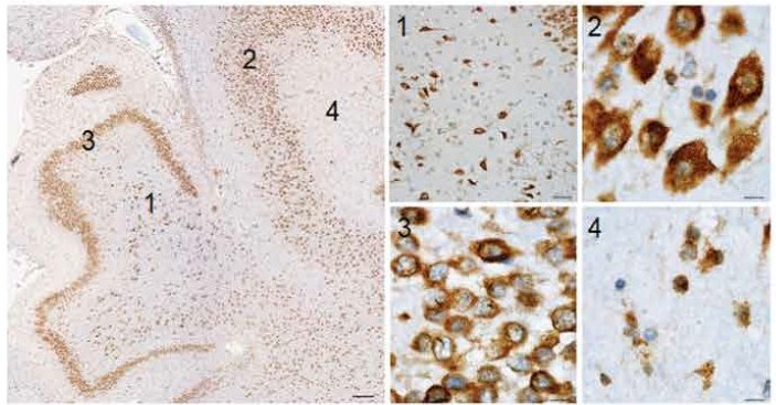
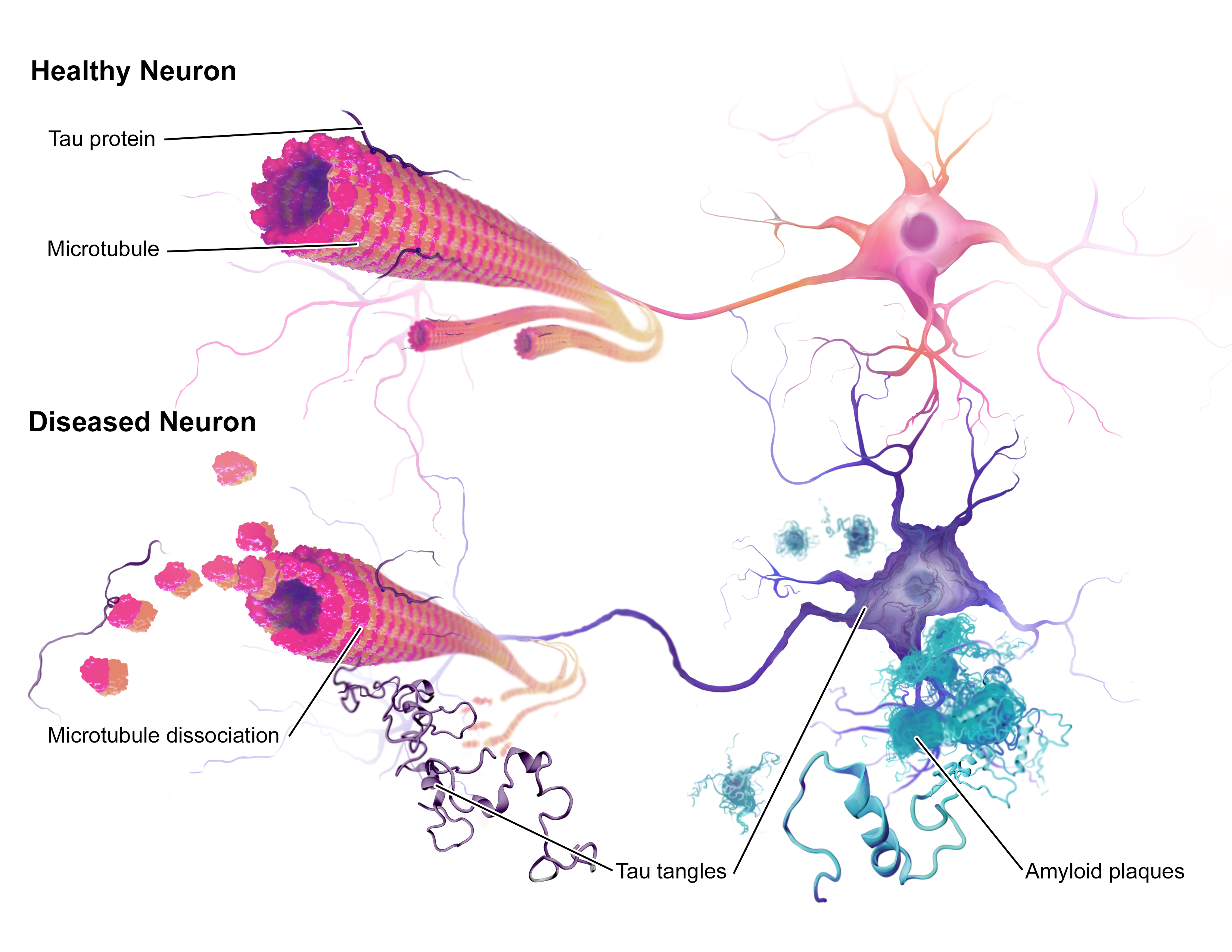
This is certainly an admirable concept given the difficulties faced thus far when trying to mimic complex diseases like Alzheimer’s in mice. Therapies that have successfully treated Alzheimer’s-like symptoms in mice have failed when trialled in humans. One reason for this could be that the mouse model of Alzheimer’s is not a close enough replica of the human disease, due to the lack of similarity between species. With non-human primates being closely related to humans, they may prove to be a better model for Alzheimer’s and other human diseases.

After this study was published it sparked discussions as to what this work means for the future of genetic cloning. The question of whether human cloning is possible was raised, to which one of the lead authors, Mu-ming Poo, responded,

Technically speaking one can clone humans…But we’re not going to do it.

The future of this work will not only likely be involved in therapeutics but in gene editing. This will allow for gene knock-out or knock-in monkey embryos with defined genetic defects for the study of a variety of diseases, allowing us to gain a better understanding of their mechanisms.

Personally, I am not sure how I feel with regards to the success of this work. On the one hand it is an astonishing achievement to have produced living non-human primate clones, given the history of unsuccessful attempts since Dolly the sheep was produced. Moreover, because of the close relation between non-human primates and ourselves with them having advanced minds as well as complex social networks, they are likely to show us more about diseases than mice ever could, particularly those affecting the brain. However, the cost of housing primates is expensive and a significant number of them would need to be produced for them to be a useful method of study. In the UK, due to the ethical restrictions surrounding primate testing, they are only used when there are no other means to address questions of importance. Therefore, current mouse models would still be sufficient in most cases, meaning that genetically uniform primates would be unnecessary. Moreover, being vegan, the treatment of animals is paramount to me. Entering into the world of research, I recognise the importance of animal models for understanding the complexity of diseases in whole organisms but I hope that one day we will be able to move away from them completely.


Drop me a comment down below about your thoughts on genetic cloning and the use of animals in research 🙂

Gum disease…not just a problem for those with a sweet tooth

A few weeks ago I was shown a really interesting paper that I just had to write about on my blog. Stephen Dominy et al., have evidence to suggest that bacteria in the mouth are related to the pathology of Alzheimer’s disease. The bacteria in question: Porphyromonas gingivalis, is the key pathogen responsible for chronic periodontitis, more commonly known as gum disease.


As mentioned in one of my previous posts, the proteins amyloid beta and tau are involved in the pathology of Alzheimer’s disease. Since the 1980’s, the leading hypothesis for the pathology of Alzheimer’s disease known as the “amyloid hypothesis”, proposes that the condition is caused by the improper function of these proteins which accumulate in the brain. This occurs outside cells in the case of amyloid-beta and inside cells for tau.

Recently gum disease and infection with P. gingivalis have been identified as significant risk factors for developing amyloid beta plaques, dementia and Alzheimer’s disease (See my introductory post for more information about Alzheimer’s disease).  One study of patients with both Alzheimer’s disease and gum disease reported decline in cognition over a 6-month period, compared to those without gum disease. This led to investigation of possible mechanisms underlying these findings. P. gingivalis produces protein-degrading enzymes known as gingipains which are essential for the survival and pathogenic capabilities of P. gingivalis. Due to the emerging evidence of P. gingivalis being involved in Alzheimer’s disease, Dominy et al., hypothesised that:

P. gingivalis infection acts in Alzheimer’s disease pathogenesis through the secretion of gingipains to promote neuronal damage.

They examined samples of Alzheimer’s disease brain tissue as well as that of neurologically normal individuals. Of the 54 Alzheimer’s disease samples taken from the hippocampus, the region of the brain important for memory, they discovered that P. gingivalis was present in over 90% of the samples. They also found a correlation between the amount of P. gingivalis in Alzheimer’s disease samples and the protein tau. Alzheimer’s disease is also associated with degradation of the grey matter of the cerebral cortex. For this reason the cerebral cortex of three Alzheimer’s disease brain samples was examined, with genetic material from P. gingivalis being detected.


Representative images of the hippocampal brain region stained for gingipains in a 63-year-old patient.

The hippocampus shows abundant gingipains in the hilus (1), CA3 pyramidal layer (2), granular cell layer (3), and molecular layer (4). High-magnification images from the indicated areas (1 to 4) exhibit a granular staining pattern consistent with P. gingivalis intracellular infection. Scale bars, 200 micrometres (overview), 50 micrometres (1), and 10 micrometres (2 to 4).

(Figure and legend modified from Figure 2: Dominy et al).

The team when on to test the cerebrospinal fluid of 10 patients diagnosed with probable Alzheimer’s disease who presented mild to moderate cognitive impairment. For these patients, P. gingivalis was present in 7 of the 10 samples. This combined with the fact that P. gingivalis associates with tau, led to them determining whether tau is a target of gingipain degradation. The results showed that the gingipains cleaved tau into fragments which have previously been found to be involved in tau tangle formation.  These findings led to the proposal that P. gingivalis does not enter the brain as a result of Alzheimer’s disease onset but it could be the cause of it. This indeed appeared to be the case when mice were infected with P. gingivalis resulting in gum disease. This led to brain infection, amyloid production, tau tangle formation and neural damage as seen in Alzheimer’s disease.

This research is vital in understanding which groups of people are more susceptible to Alzheimer’s disease, one of which is individuals with Down syndrome. Within this group there is a high prevalence of dementia with Alzheimer’s-type pathology. This is believed to be due to the fact that the amyloid precursor protein gene which gives rise to amyloid beta is present on chromosome 21 and this chromosome is triplicated in Down syndrome. The team point out that the occurance of P. gingivalis is found to be significant in the mouths of Down syndrome patients beginning in early childhood compared to age-matched individuals without Down syndrome, indicating that P. gingivalis is present at abnormal levels in Down syndrome patients. The susceptibility of Down syndrome patients to P. gingivalis is not yet clear but is suggested to be due to having weakened immune systems.

Future work will need to identify the route of entry into the brain by P. gingivalis. However, the group does propose that once in the brain it spreads from one neuron to the next, which may be the cause of the tau pathology in Alzheimer’s diseased brains, which had already been suggested to occur in this manner. Casey Lynch a member of the team describes their hypothesis that P. gingivalis infection is the cause rather than a result of Alzheimer’s disease as being,

A universal hypothesis of pathogenesis, fully explaining the cause of Alzheimer’s disease.

This paper was certainly very exciting to read. The concept of the “amyloid hypothesis” is well-supported within the scientific community. For this new study to propose that amyloid beta and/or tau dysfunction is not the cause but one of the effects of the onset of Alzheimer’s disease, is truly ground-breaking.

In the latter part of their paper they describe methods for blocking gingipain activity. This involved administering small-molecular inhibitors to mice with Alzheimer’s disease pathology resulting from P. gingivalis infection. The results of which showed reduced infection, prevention of amyloid production and lowered inflammation. The small-molecular inhibitors are currently being trialled in human clinical studies for Alzheimer’s disease. If these studies have positive outcomes then this group may have just found the cause and cure of Alzheimer’s disease!

Hello and Welcome!

My name is Megan, I am a fourth-year MSci Biochemistry and Genetics student at the University of Nottingham. This blog will serve as an online journal during my fourth-year project. I will review current science papers in the field as well as giving my own opinions on popular science. Stay tuned, there will be some exciting posts to follow!


My main scientific interest is in neurodegenerative disease, particularly Alzheimer’s disease and how cellular metabolism is implicated. Alzheimer’s disease is the leading cause of dementia in the elderly. It is associated with progressive decline of brain function which can affect memory and cognitive function. With an ageing population it is important for us to understand how this disease materialises and ways in which it can be treated.

There are two proteins that largely contribute to the pathology of the disease known as, amyloid beta and tau. The role of tau proteins is to stabilise microtubules within the distal region of neuronal axons.  When the activity of tau kinases acting on tau is increased, the tau proteins misfold and aggregate forming neurofibrillary tangles inside neurons and less commonly in glial cells.  Such tangles block nerve synapses and prevent nutrient transport between cells, causing neuronal death. The normal function of amyloid beta is not well understood. Pathological amyloid beta forms aggregates called amyloid fibres which are the main component of amyloid plaques, the pathological hallmark of Alzheimer’s disease. Unlike tau neurofibrillary tangles, the build-up of amyloid beta within amyloid plaques occurs outside the neurons.


An illustration of the neuropathology of Alzheimer’s disease.

(Taken from: BruceBlaus [CC BY-SA 4.0 (https://creativecommons.org/licenses/by-sa/4.0)])

My fourth-year project involves using astrocytes which are a type of glial cell. Astrocytes are star shaped cells distributed throughout the brain and spinal cord supporting neurons within the central nervous system. Astrocytes connect neurons to the blood brain barrier and therefore serve as a mediator for transport of nutrients/molecules for signalling and overall neuron function. It was originally believed that they were merely supporting cells and while this is their primary function, other roles have come to light. For this reason, we are interested in whether they play a part in the pathology and progression of Alzheimer’s disease. Therefore, I am investigating the effects of amyloid beta on the metabolism and morphology of astrocytes.

Previous work by Garwood et al., showed that the level of interleukin 6 (IL-6) secretion from astrocytes was increased when incubated with amyloid beta. IL-6 is a cytokine known to activate the signalling molecule STAT3 through binding to its receptor on the surface of cells. This results in activation of the JAK-STAT pathway and modulation of STAT3 target genes in the nucleus.


Simplistic diagram of the JAK-STAT signalling pathway

(Taken from: Pharmstudice [CC BY-SA 4.0 (https://creativecommons.org/licenses/by-sa/4.0)])

During my project I am aiming to elucidate whether the incubation of astrocytes with amyloid beta will affect the signalling of STAT3 and whether this is likely to be due to the interplay of IL-6 and STAT3. My project will begin to address whether STAT3 signalling in astrocytes plays a role in Alzheimer’s disease and whether it affects the functioning of neurons.

My project began with optimising a method called immunocytochemistry (ICC). This involves using antibodies to stain specific proteins in cells for visualisation under a microscope. During the autumn term and beginning of the spring term I optimised ICC for the proteins STAT3 and GFAP. GFAP is a structural protein present in astrocytes and serves as a marker for the cells. I began the process optimising use of the fluorophore Alexa-488, a green fluorescent marker attached to an antibody. Although this worked, the amyloid beta I will be using has the same fluorophore attached to it, so I went on to optimise other antibodies. Next was Alexa-568, a red marker, which worked well for both STAT3 and GFAP. I then tried Alexa-405, a blue marker, however, it didn’t work after two attempts. This means that because STAT3 and GFAP worked well with the same antibody I will have to incubate my cells with amyloid beta, staining for STAT3 and GFAP separately instead of both at the same time with amyloid beta- Below is a gallery of some of the conditions I optimised during my preliminary work, click them to view them in full.


Now that the preliminary work is completed, I shall be moving on to the investigative portion of my project. First, I will be incubating my cells with the amyloid beta and performing ICC for STAT3 and GFAP with the conditions I determined before. This will be followed by analysis of my images which will involve:

  • Counting the frequency of amyloid beta uptake
  • Measuring the size of amyloid beta accumulation
  • Measuring the length of cell filopodia (extensions from the cell body)
  • Measuring the amount of fluorescence for STAT3 and GFAP

After this I will do an MTT assay and a glucose uptake assay. The MTT assay is a technique used to determine cell viability and the glucose uptake assay is a method for determining the metabolic activity of a cell.

Should STAT3 be affected by amyloid beta incubation and if there is enough time towards the end of my project, I would like to run an ELISA assay on the media that the astrocytes have been incubated in to see if IL-6 is present. If IL-6 is present, it may be the cause of alterations in STAT3 activity. If STAT3 is not affected by amyloid beta, an ELISA assay for a variety of cytokines could be used to determine alterations in the levels of other cytokines as a result of amyloid beta uptake.


That just about sums up my work so far and my plans for my project. If you have any questions or want to know more drop me a comment below 🙂